Resource Library
We believe health starts with knowledge and information. Explore our resources on all things related to common sports medicine conditions, preventative techniques, and more.
We believe health starts with knowledge and information. Explore our resources on all things related to common sports medicine conditions, preventative techniques, and more.
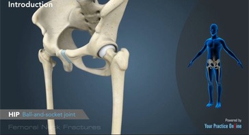
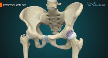
The hip is a ball-and-socket joint made up of the head of the thigh bone or femur that acts as the ball and fits into the rounded socket of the hip bone or acetabulum.

The hip joint is one of the body's largest weight-bearing joints and is the point where the thigh bone (femur) and the pelvis (acetabulum) join.
The hip joint is also known as the ball and socket joint, where the ball (femoral head) of the thigh bone fits into the socket (acetabulum) of the pelvis bone.
The hip joint is the largest weight-bearing joint in the human body. It is also referred to as a ball and socket joint and is surrounded by muscles, ligaments, and tendons.
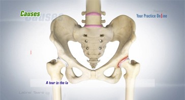
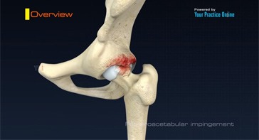
A hip labral tear is an injury to the labrum, the cartilage that surrounds the outside rim of your hip joint socket.

Slipped capital femoral epiphysis (SCFE) is a common hip disorder in adolescents causing slippage or separation of the femoral head (ball at the upper end of the femur bone)...
Hip synovitis, also called transient hip synovitis or toxic synovitis is a condition in which there is inflammation of the synovial tissues surrounding...
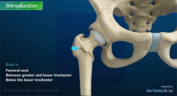
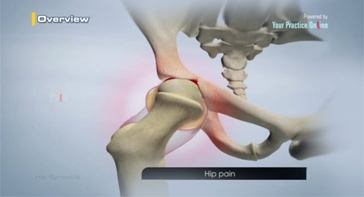
A hip fracture is a break that occurs near the hip in the upper part of the femur or thigh bone. The thigh bone has two bony processes on the upper part...
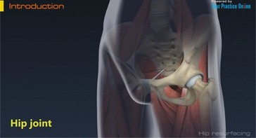
The hip joint is also known as a ball and socket joint, where the ball (femoral head) of the thigh bone fits into the socket (acetabulum) of the pelvis bone.
Posterior hip replacement is a minimally invasive hip surgery performed to replace the hip joint.
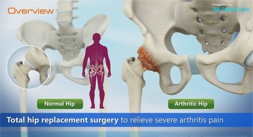
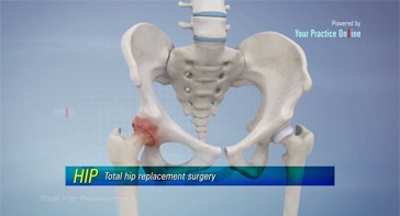
Total hip replacement surgery is an option to relieve severe arthritis pain that limits your daily activities.
The hip joint is one of the body's largest weight-bearing joints, located between the thigh bone (femur) and the pelvis (acetabulum).
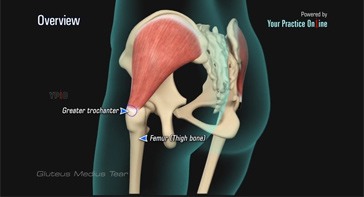
The gluteus medius is one of the major muscles of the hip and is essential for movement of the lower body and keeping the pelvis level during ambulation.
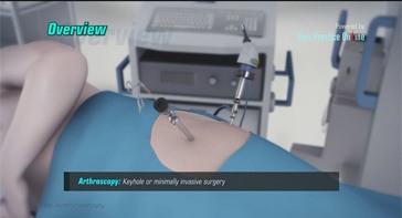
Arthroscopy, also referred to as keyhole or minimally invasive surgery, is a procedure in which an arthroscope is inserted into a joint to check for...
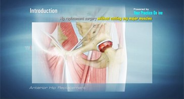
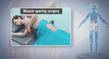
Anterior hip replacement is a minimally invasive hip surgery performed to replace the hip joint without cutting through any muscles.
The hip joint is one of the largest weight-bearing joints in the body, formed by the thigh bone or femur and the acetabulum of the pelvis.
An osteotomy is a surgical procedure that involves cutting and reshaping of a bone.
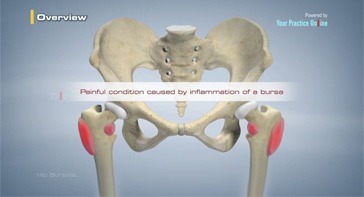
Hip bursitis is a painful condition caused by inflammation of a bursa in the hip.
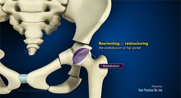
Periacetabular osteotomy is a surgical procedure to treat a congenital hip condition called hip dysplasia.
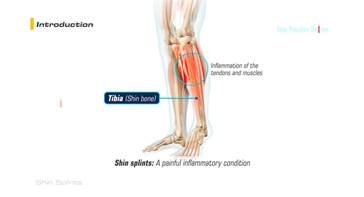
Shin splints is a painful condition caused by inflammation of the tendons and muscles of thetibia or shin bone.
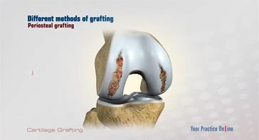
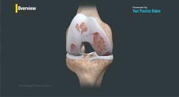
Cartilage grafting is a surgical procedure that replaces damaged cartilage with healthy cartilage from a nonâ€weight bearing joint.
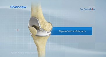
Total knee replacement is a very successful surgical treatment for knee arthritis. Over the years, minimally invasive surgical techniques...
Patellofemoral pain syndrome, also referred to as PFPS, is one of the most commonly reported knee problems, accounting for one in four knee complaints seen by Orthopaedists.
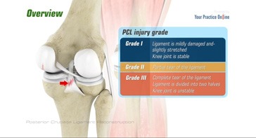
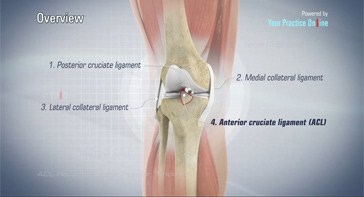
Posterior cruciate ligament (PCL), one of four major ligaments of the knee, is situated at the back of the knee.
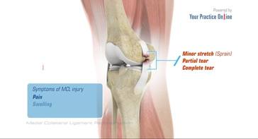
Medial collateral ligament (MCL) is one of four major ligaments of the knee that connects the femur (thigh bone) to the tibia...
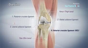
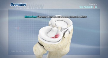
Anterior cruciate ligament is one of the four major ligaments of the knee that connects the femur (thigh bone) to the tibia (shin bone) and helps stabilize your knee joint.
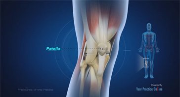
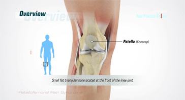
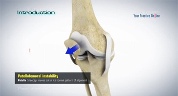
The patella or kneecap is a small bone present in the front of your knee where the thighbone meets the shinbone.
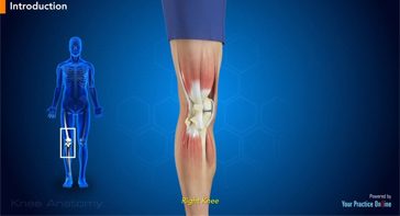
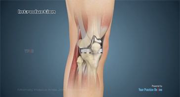
The knee is a complex joint made up of different structures including bones, tendons, ligaments and muscles.
The knee is made up of the femur (thigh bone), the tibia (shin bone), and patella (kneecap).
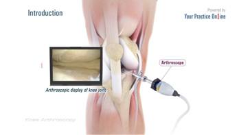
Knee Arthroscopy is a common surgical procedure performed using an arthroscope, a viewing instrument, to look into the knee joint to diagnose or treat a knee problem.
The meniscus is a small, "C" shaped piece of cartilage in the knee joint. Each knee has two menisci, the medial meniscus on the inner aspect...
Anterior cruciate ligament is one of the four major ligaments of the knee that connects the femur (thigh bone) to the tibia (shin bone) and helps stabilize the knee joint.
Knee pain and stiffness can be disabling and difficult to treat. It can limit an individual’s lifestyle and negatively impact body image and emotional well-being.
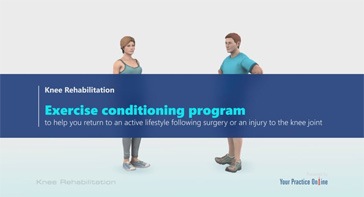
Knee Rehabilitation is an exercise conditioning program to help you return to an active lifestyle following surgery or an injury to the knee joint.
The knee can be divided into three compartments: patellofemoral, the compartment in front of the knee between the knee cap...
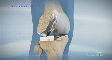
The knee can be divided into three compartments: patellofemoral, medial and lateral compartment.
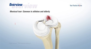
The meniscus is a C-shaped cartilage ring that acts like a cushion between the shinbone and the thighbone.
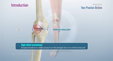
High tibial osteotomy is a surgical procedure to relieve pressure on the damaged site of an arthritic knee joint.
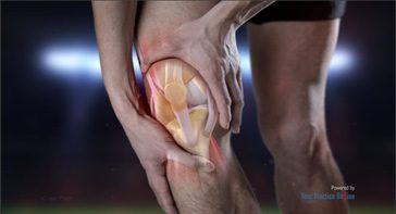
Knee pain is a common condition affecting individuals of various age groups. It not only affects movement, but also impacts quality of life.
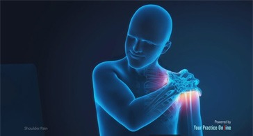
Shoulder pain is a common orthopedic condition with pain ranging from mild to unbearable. You may experience pain with movement of the shoulder or at rest.
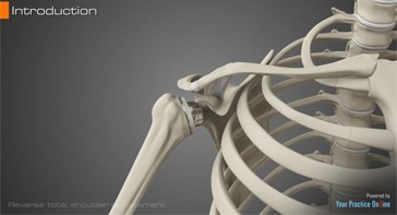
Reverse total shoulder replacement, is an advanced surgical technique specifically designed for rotator cuff tear arthropathy...
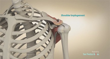
A subacromial decompression is a surgery performed to treat shoulder impingement, one of the most common causes of shoulder pain.
The shoulder joint is a ball and socket joint. A 'ball' at the top of the upper arm bone (the humerus) fits neatly into a 'socket', called the glenoid...
The hip joint is also known as the ball and socket joint, where the ball (femoral head) of the thigh bone fits into the socket (acetabulum) of the pelvis bone.
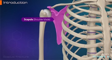
The shoulder is made up of the clavicle (collar bone), humerus (upper arm) and scapula (shoulder blade).
The shoulder is a ball and socket joint made up of the scapula (shoulder blade), clavicle (collarbone), and humerus (upper arm bone).
Shoulder impingement is the inflammation of the tendons of the shoulder joint. It is one of the most common causes of pain in the shoulder.
The upper arm is made up of the humerus bone. The head of the humerus fits into a shallow socket in your scapula (shoulder blade) to form the shoulder joint.
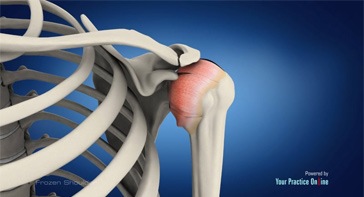
Frozen shoulder, also called adhesive capsulitis, is a condition in which you experience pain and stiffness in your shoulder.
The shoulder is a ball and socket joint where the ball is formed by the head of the upper arm bone or humerus and the socket is formed by a shallow cavity...
The shoulder joint (glenohumeral joint) is a ball and socket joint, where the head of the upper arm bone (humerus) attaches to the shoulder socket (glenoid cavity).
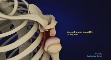
The shoulder joint provides a wide range of movement to the upper extremity, but injuries or repeated dislocations can cause instability to the joint.
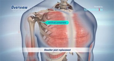
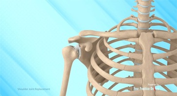
Shoulder joint replacement is a surgical procedure to replace damaged bone surfaces with artificial components to relieve pain and improve functional ability in the shoulder joint.
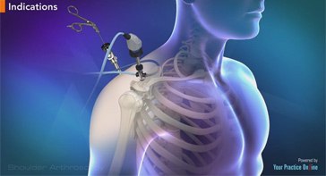
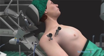
Arthroscopy is a minimally invasive diagnostic and surgical procedure performed for joint problems.
The shoulder is a ball and socket joint made up of three bones, namely the humerus, scapula, and clavicle.
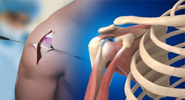
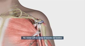
Arthroscopic rotator cuff repair is a minimally invasive surgery to repair an injured or torn rotator cuff using an arthroscope.
The shoulder is considered one of the largest joints in the body, formed by the articulation of the shoulder blade (scapula) and bone of the upper arm (humerus).
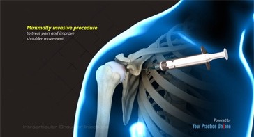
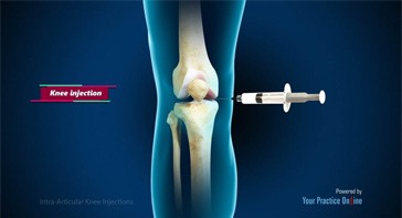
An intraarticular shoulder injection is a minimally invasive procedure to treat pain and improve shoulder movement.
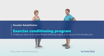
Shoulder Rehabilitation is an exercise conditioning program to help you return to an active lifestyle following surgery or an injury to the shoulder joint.
Your shoulder joint is a ball and socket joint made up of the upper arm bone, the shoulder blade and the collarbone.
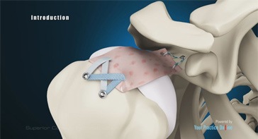
Superior capsular reconstruction is a surgical procedure performed to restore shoulder stability in irreparable rotator cuff tears.
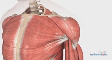
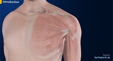
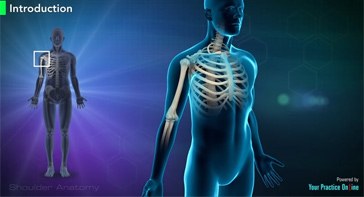
The shoulder is a complex joint where several bones, muscles, and ligaments connect the upper extremity to the chest.
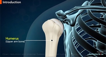
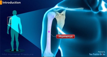
The humerus is the upper arm bone. A fracture of the proximal humerus, the region closest to the shoulder joint, can affect your work and activities of daily living.
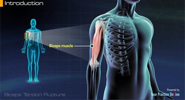
The biceps muscle is present on the front side of your upper arm and functions to help you bend and rotate your arm.
The shoulder is a highly movable body joint that allows various movements of the arm. It is a ball-and-socket joint, where the head of the humerus (upper arm bone)...
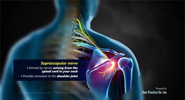
A suprascapular nerve block is a procedure to inject an anesthetic or a numbing medication along with an antiinflammatory in the region of the suprascapular nerve...
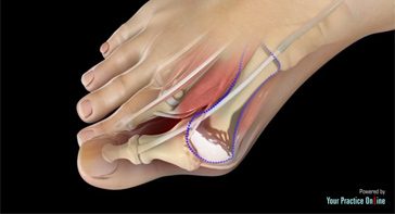
A bunion, also known as hallux valgus, is bony prominence at the base of the big toe, which often results in pain, redness and rubbing in footwear.

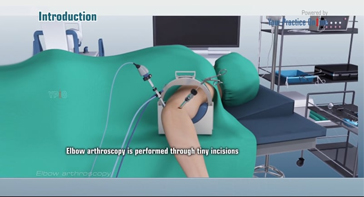
Ankle arthroscopy is a minimally invasive surgical procedure in which an arthroscope, a small, soft, flexible tube with a light and video camera at the end...
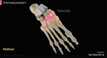
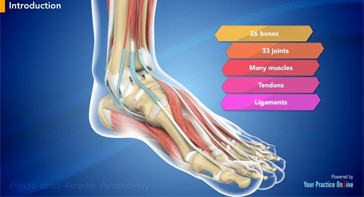
The foot has 26 bones, and can be divided into 3 parts:
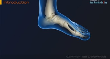
Anatomically the foot is divided into the forefoot, mid foot and hind foot. The forefoot consists of your toe bones, called phalanges, and metatarsal bones...
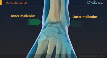
The ankle joint is composed of three bones: the tibia, fibula, and talus which are articulated together.
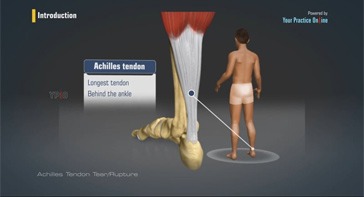
Tendons are soft tissues connecting muscles to bone. The achilles tendon is the longest tendon in the body and is present behind the ankle, joining the calf muscles...
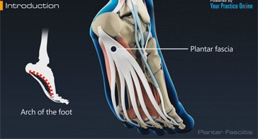
The plantar fascia is a long, thin ligament present along the bottom of the foot that creates the arch of the foot.
The ankle is composed of bones forming a joint and ligaments are the elastic structures which are responsible for holding these bones in their proper place.
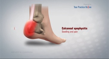
Sever’s disease is a painful inflammation of the growth plate in the heel. Growth plates are areas at the end of children’s bones that undergo changes so bone growth can occur.
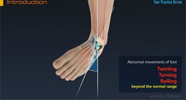
The foot and ankle is a complex joint involved in movement and providing stability and balance to the body.
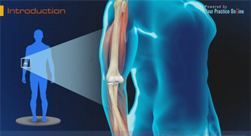
The elbow is a complex joint formed by the articulation of three bones – the humerus, radius and ulna. The elbow joint helps
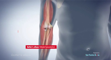
Golfer’s elbow, also called Medial Epicondylitis, is a painful condition that occurs due to repeated muscle contractions in the forearm
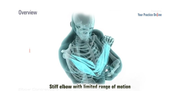
Elbow contracture refers to a stiff elbow with limited range of motion. It is a common complication following elbow surgery, fractures, dislocations, and burns.
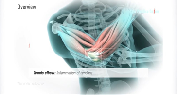
Tennis elbow, also known as lateral epicondylitis, is a condition characterized by elbow pain due to overuse or overstretching of the elbow.
The elbow is a complex hinge joint formed by the articulation of three bones- humerus, radius, and ulna.
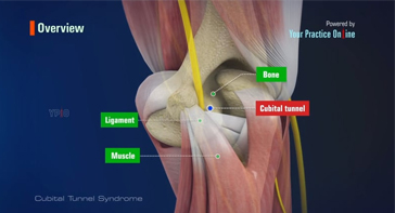
Cubital Tunnel Syndrome is a condition characterized by compression of the ulnar nerve in an area of the elbow called the cubital tunnel.
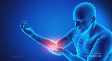
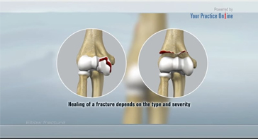
Elbow pain is a common condition that can have various causes. The pain can range from a burning sensation to a sharp pain.
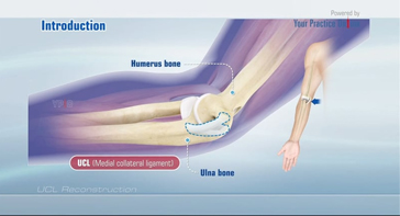
The UCL, also called medial collateral ligament, is located on the inside of the elbow and connects the ulna bone to the humerus bone.
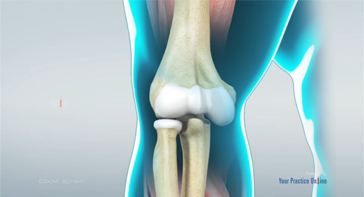
The Elbow is a complex hinge joint formed by the articulation of three bones - humerus, radius and ulna.
The Elbow is a complex hinge joint formed by the articulation of three bones - humerus, radius and ulna. The upper arm bone
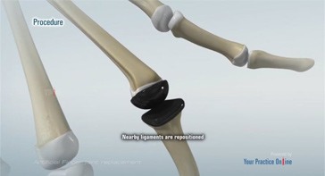
Elbow Joint Replacement, also referred to as Total Elbow Arthroplasty is an operative procedure to treat the symptoms of arthritis
The hands are made up of 27 bones, which are grouped into carpals, metacarpals and phalanges. Each bone is separated by the articular cartilage...
The ability to bend the fingers is governed by supportive tendons that connect muscles to the bones of the fingers.
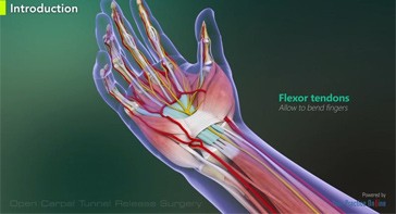
The carpal tunnel is a narrow passage on the palm side of your wrist. Small wrist bones known as carpals form the bottom and sides of your carpal tunnel...
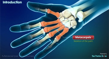
Metacarpal fracture is a condition characterized by the breakage or dislocation of the long hand bones called metacarpals that form the skeleton of the palm.
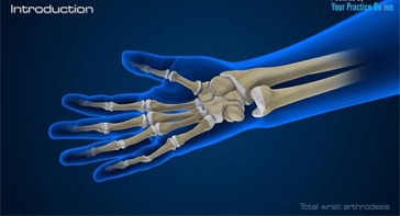
The wrist joint is one of the most complex joints in the human body. Numerous joints and bones contribute to the strength and stability of the wrist.
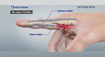
Tendons are the bands of fibrous connective tissue that connect muscles to bone. Tendons aid in movement of the fingers, hand and all other body parts.
The hands are made up of 27 bones, which are grouped into carpals, metacarpals and phalanges.
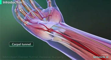
The carpal tunnel is a narrow passageway on the palm side of your wrist. Small wrist bones known as carpals form the bottom and sides of your...
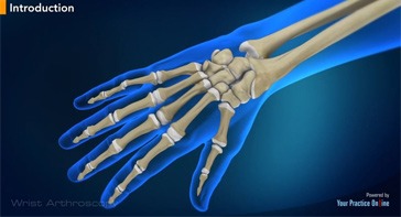
Your wrist is a complex joint made up of eight small bones called carpal bones. These bones are supported by connecting ligaments.

The hand is made up of 27 bones that form the wrist, palm, and fingers. Fingers can easily injure from daily activities, and fractures are common injuries that can occur.
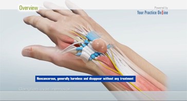
Ganglion cysts are fluid-filled lumps that most commonly develop along the tendons or joints of wrists or hands.
Dupuytren’s Contracture is a hand condition where thickening of the underlying fibrous tissues of the palm cause the fingers to bend inward.
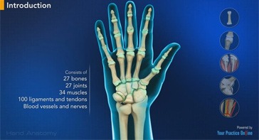
The human hand is made up of the wrist, palm, and fingers and consists of 27 bones, 27 joints, 34 muscles, over 100 ligaments and tendons, and many blood vessels and nerves.
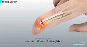
Mallet finger is a condition where the end of the finger is bent and does not straighten.
The wrist is comprised of two bones in the forearm, the radius and ulna, and eight tiny carpal bones in the palm.
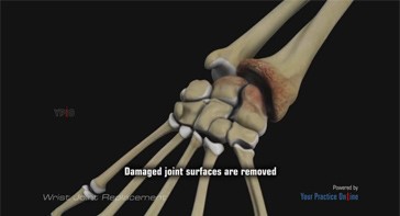
The wrist is a complex joint made up of 8 carpal bones aligned in two rows with four bones present in each row.
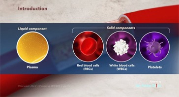
PRP is a relatively new method of treatment for several orthopaedic conditions such as muscle, ligament, and tendon injuries; arthritis; and fractures.
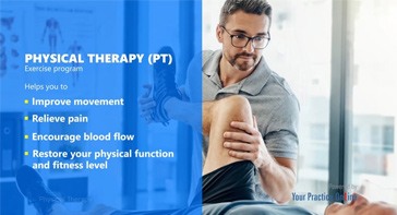
Physical Therapy, often referred to as PT, is an exercise program that helps you to improve movement, relieve pain, encourage blood flow for faster healing...
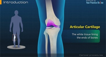
Articular Cartilage is the white tissue lining the ends of bones where they connect to form joints.
Articular cartilage is the smooth, shiny, white tissue covering the ends of bones those form a joint.
Cartilage grafting is a surgical procedure that replaces damaged cartilage with healthy cartilage from a nonâ€weight bearing joint.
Anterior cruciate ligament is one of the four major ligaments of the knee that connects the femur (thigh bone) to the tibia (shin bone) and helps stabilize the knee joint.
Get in touch with our team so we can put together a customized plan to get you back to your best and strongest self.